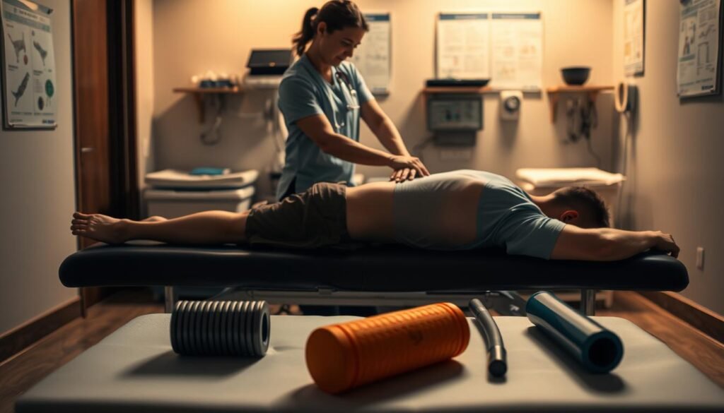
Surprising fact: up to 80% of people with acute low back pain feel a big improvement within 30 days.
This guide explains what a lumbosacral strain is and why the lower back can become sore after heavy lifting, twisting, or sports. A lumbar strain is an injury to the low back’s muscles and tendons. It often causes spasms and aching that limit daily activity.
The article shows practical first-line management you can use now: early gentle movement, heat or ice, short-term NSAIDs, and guided exercises. Imaging is not usually needed unless red flags like fever, spreading leg pain, or bladder changes appear.
Readers will learn stage-based treatments and simple checks for when to see healthcare. The goal is faster recovery, less back pain, and smarter ways to lower the risk of repeat injuries.
Key Takeaways
- Most acute low back pain improves substantially within about 30 days.
- Early movement, heat/ice, and short-term meds are common first-line treatments.
- Avoid imaging unless red flags or serious symptoms are present.
- Know warning signs—fever, leg weakness, spreading pain, or bladder changes.
- Stage-based management helps you return to normal activities safely.
What Is a Lumbosacral Strain?
A soft-tissue injury in the lumbar area causes focal back pain and often limits movement. These injuries affect muscles, tendons, and ligaments that support the lower spine. The result is localized soreness, tenderness, and sometimes protective muscle spasms.
How lumbar muscles, tendons, and ligaments get injured
When forces exceed what the supporting structures can handle, fibers can overstretch or tear. Sudden rotation, full flexion, or heavy load commonly triggers this.
Spasm follows when a muscle contracts persistently. That reduces blood flow and lets waste products build up, which increases pain.
Common activities that trigger low back strain
Everyday tasks and sports both cause problems. Twisting while lifting boxes, bending during cleaning, and high-torque moves in weightlifting or football often precipitate injury.
Tight hamstrings, weak abdominals, and exaggerated lumbar curve raise the risk by placing structures at a mechanical disadvantage.
Quick comparison
| Trigger | Example | Result |
|---|---|---|
| Twisting | Golf swing, reaching awkwardly | Muscle micro-tears, localized back pain |
| Heavy lifting | Moving furniture, deadlifts | Tendon overload, spasms |
| Repetition | Yard work, repetitive bending | Fatigue, increased injury risk |
Who’s at Risk: Causes and Risk Factors for Lower Back Strain
A mix of posture, repetitive loading, and lifestyle choices determines who is more likely to hurt their lower back. Recognizing these patterns helps prevent injury and speed recovery.
Posture, core weakness, and tight hamstrings
Poor posture and weak abdominal muscles shift load onto the lumbar muscles. That increases the chance of a soft tissue strain during routine tasks.
Tight hamstrings tilt the pelvis forward and raise lumbar curve, concentrating stress when you bend or lift.
Repetitive or heavy lifting and rotated positions
Repetitive or heavy lifting, especially with the trunk rotated or flexed, is a leading cause of lower back injuries. These positions reduce the spine’s mechanical advantage and trigger muscle overload.
Lifestyle and psychosocial factors
Smoking, high stress, low job control, and long periods of sitting also increase risk. Psychosocial “yellow flags” predict pain that lasts beyond expected weeks.
- Improve ergonomics and body mechanics.
- Build core strength and hamstring mobility.
- Address stress and workplace demands early.
| Risk factor | How it raises risk | Simple fix |
|---|---|---|
| Poor posture | Shifts load to lumbar muscles | Postural exercises, ergonomic chair |
| Repetitive lifting | Fatigues stabilizing tissues | Use safe lifting technique, breaks |
| Smoking / stress | Slows healing, increases perceived pain | Quit support, stress reduction |
For related spine issues and management options, see dolor facetario for additional context.
Symptoms to Recognize vs. Red Flags You Shouldn’t Ignore
Most episodes of sudden back pain are self-limited, but a few warning signs need prompt attention. The next sections help you tell normal recovery symptoms apart from problems that require medical evaluation.
Typical symptoms of a lumbar muscle injury
Common signs include sudden low back pain, soreness when you press over the muscles, and painful spasms that worsen with movement.
Discomfort usually stays in the lower back rather than shooting below the knee. Gentle rest and position changes often ease symptoms.
Neurologic and systemic red flags
Watch closely for symptoms that suggest a more serious condition.
- Pain spreading down the leg or new numbness/tingling in the foot.
- New leg weakness or difficulty walking.
- Fever, chills, or unexplained weight loss.
- Loss of bladder or bowel control or saddle numbness — seek emergency care.
“Early recognition of red flags lets clinicians rule out infection, fracture, cancer, or cauda equina and start appropriate care.”
When to call a healthcare provider in the United States
Contact a clinician promptly if pain is constant, severe at rest, or not improving after several days to a week.
Providers may order blood tests (CBC, ESR, CRP) or urgent imaging when infection or inflammatory conditions are a concern.
| Finding | Likely meaning | Typical action |
|---|---|---|
| Fever + worsening back pain | Possible infection | Blood tests, urgent imaging, antibiotics |
| Leg weakness or numbness | Neurologic compression | Neurologic exam, MRI, urgent referral |
| Pain not easing after 7 days | Unusual course | Clinical review, consider further testing |
When unsure, err on the side of caution. For guidance on timing and red flags, see when to worry about lower back.
How Lumbosacral Strain Is Diagnosed
Diagnosis begins with a focused conversation about recent activity, pain patterns, and how symptoms limit daily life.
History and focused physical exam
Clinicians ask when and how the injury happened, what movements provoke pain, and whether any numbness or leg symptoms exist.
The physical exam inspects posture and gait, checks lumbar range of motion, and palpates paraspinal muscles for localized tenderness.
Strength, reflexes, and sensation are usually normal in a pure lumbar strain.
A straight leg raise may reproduce back pain but should not cause radicular pain below the knee.
Imaging and laboratory testing
Imaging (X-ray, CT, MRI) adds little for uncomplicated acute back pain and is reserved for red flags or when symptoms persist beyond about six weeks.
Blood tests (CBC, ESR, CRP) are used only if infection or inflammatory conditions are suspected based on history or exam.
Ruling out other sources of pain
Because hip or sacroiliac joint problems can mimic a lumbar injury, clinicians use targeted maneuvers (FABER, compression, distraction, thigh thrust, Fortin’s finger, Gaenslen’s) to pinpoint the source.
Practical point: a clinical diagnosis guides conservative treatment and helps avoid unnecessary tests. Shared decision-making with your healthcare provider ensures tests are used when they will change management.
| Assessment step | Key findings | Usual action |
|---|---|---|
| History | Onset, provocation, function limits | Guide exam and need for urgent care |
| Physical exam | Paraspinal tenderness, limited ROM, normal neuro exam | Conservative treatment, targeted rehab |
| Imaging / labs | Red flags, persistent pain, fever, weakness | X-ray/CT/MRI or CBC/ESR/CRP as indicated |
| Hip / SI testing | Reproduced pain with pelvic maneuvers | Refer or target treatment to hip/SI structures |
For practical guidance on diagnosis and early management, see diagnosis and early care for lumbar.
Course of Recovery: Acute, Subacute, and Chronic Low Back Pain
Most people notice the worst back pain within the first hours to days, then enter a period of steady recovery over the coming weeks.
Acute (0–4 weeks): Pain tends to be sharpest early on and often improves as inflammation settles. About 90% of acute cases show clear gains by roughly 30 days with sensible activity and care.
Subacute (4–12 weeks): The focus shifts to rebuilding capacity. Gradual increases in walking and core work protect the lumbar area and reduce the risk of recurrence.
Chronic (>12 weeks): When symptoms persist past 12 weeks, a comprehensive plan that mixes physical conditioning with psychological support often helps more than simple rest.
Too much rest early can worsen deconditioning. Gentle movement, short frequent walks, and staged exercises promote blood flow and ease stiffness without overloading the back.
- Expect brief flare-ups when starting new exercises; small progressions are safer.
- Set clear short-term goals — today, this week, this month — to track recovery and maintain motivation.
- Watch for yellow flags (high disability, low mood, poor support) and address them early to reduce risk of chronic symptoms.
For evidence on recovery timelines and predictors of persistent pain, see a clinical review at recovery and prognosis. For practical timelines on healing and activity progression, this resource on disc healing offers useful comparisons: how long to heal.
Lumbosacral Strain: Evidence-Based Treatments by Stage
Managing lower back pain means matching treatments to the recovery phase, from immediate relief to long-term conditioning.
Acute phase (0–4 weeks)
Stay active within comfort and avoid prolonged bed rest beyond a day or two. Use superficial heat or ice and simple compression for short-term comfort. Consider NSAIDs for modest short-term pain relief; acetaminophen is less helpful, and opioids are not recommended. Short-term muscle relaxants may ease spasms but can cause drowsiness, so use them cautiously.

Progress to graded activity and structured physical therapy. Emphasize core stabilization and gradual loading to restore lumbar muscle endurance. Routine imaging remains unnecessary unless red flags or poor progress appear after several weeks of guided care.
Chronic phase (>12 weeks)
Adopt a multidisciplinary plan that blends exercise therapy with cognitive behavioral therapy or mindfulness to reduce pain-related disability. Consider sustainable adjuncts such as tai chi, yoga, acupuncture, or spinal manipulation where evidence supports benefit.
What major guidelines recommend
| Guideline | Key stance | Clinical note |
|---|---|---|
| ACP / JOSPT | Noninvasive care first | Prioritize activity, PT, and education |
| VA/DoD (2022) | Limit opioids; NSAIDs with caution | Supports short-term NSAIDs; avoids opioids |
| WHO | Conditional use of meds | Avoid routine muscle relaxants in older adults |
Practical point: Across stages, the mainstay is progressive movement and tailored rehabilitation. Work with your healthcare team to choose treatments that fit your goals, history, and daily demands.
Home Management: Pain Relief, Rest, Heat and Ice, and Compression
Practical steps at home can calm inflammation and reduce painful muscle spasms. Early, simple care speeds recovery and cuts the chance of a longer episode.
Timing heat vs. ice and safe application at home
Cold reduces swelling in the first 48–72 days after a flare; apply ice for 15–20 minutes at a time. Use a thin towel between skin and ice to prevent frostbite.
After the initial days, use short bouts of heat (about 15 minutes) to relax tight tissue. Never sleep on a heating pad and always protect skin with a barrier.
OTC medications and when to avoid prolonged bed rest
Over-the-counter NSAIDs like ibuprofen can lower pain and inflammation. Follow label directions and check with a clinician if you use blood thinners, have ulcers, or kidney disease.
Short periods of rest are fine, but prolonged bed rest weakens muscle and stiffens joints. Gentle walking and safe body mechanics (bend at hips/knees, keep loads close) usually help more than staying still.
| Home measure | When to use | Quick tip |
|---|---|---|
| Ice | First few days | 15–20 min, towel barrier |
| Heat | After initial swelling eases | 15 min, never overnight |
| Compression | During activity transitions | Supportive, snug but not restrictive |
Exercises for Strength and Flexibility
Improving control around the pelvis and hips reduces load on the lower back during daily tasks. A brief, steady program that blends core work and mobility helps ease pain and restore function.

Core stabilization and graded lumbar strengthening
Start with abdominal bracing, pelvic tilts, and bridge holds to build strength that supports the spine. These moves unload the back and teach safe muscle activation.
Progress to bird-dog, side planks, and hip hinges. Increase hold time or sets week by week to add load without flaring symptoms.
Stretch hips and hamstrings
Gentle hip and hamstring stretches reduce anterior pelvic tilt and protect the lumbar region during sitting and lifting. Hold each stretch briefly and avoid bouncing.
Movement options and safe progress
Low-impact activities—walking, tai chi, Pilates, and yoga—improve flexibility, coordination, and confidence without overloading healing muscles.
- Increase one variable at a time: reps, sets, or resistance.
- Allow 24–48 hours between harder sessions and expect mild muscle soreness.
- Scale back if pain becomes sharp, radiates, or causes new weakness—see a physical therapist for tailored progression.
Prevention, Ergonomics, and Return to Work or Sports
Good ergonomics and graded activity cut the chance of recurring back problems and speed safe returns to work or sport.
Body mechanics for lifting and daily activities
Lift with hips and knees. Bend at your hips and knees, keep a neutral spine, and hold the load close to your body. Avoid twisting while carrying items to protect muscles and tendons.
Use your legs as the power source. This lowers force on the lumbar area and reduces the risk of re-injury.
Workplace setup and early return-to-work strategies
Early return to work with temporary adjustments improves long-term outcomes. Prolonged absence raises the risk of not returning: about 50% at six months and under 10% at one year.
- Adjust monitor height, keep keyboard and mouse within easy reach, and use lumbar support.
- Plan micro-breaks every 30–60 minutes to stand, walk, and mobilize the back.
- Negotiate light duties, task rotation, and extra breaks while recovering to stay engaged and safe.
Sport-specific tips and graded return-to-activity
Rebuild fundamentals first: core strength, hip mobility, and correct technique. Add sport-specific loads slowly and monitor for pain.
Use a stepwise plan: increase volume or intensity in stages and confirm tolerance before advancing. Functional tests like the Back Performance Scale and the Biering-Sorensen test help gauge readiness.
| Focus | Action | Why it helps |
|---|---|---|
| Safe lifting | Hip-knee bend, neutral spine, keep load close | Reduces lumbar load and protects tendons and muscles |
| Workstation | Screen at eye level, feet flat, lumbar support | Maintains natural spine curves and lowers daily fatigue |
| Return-to-work | Light duties, breaks, gradual load increase | Speeds recovery and lowers long-term disability risk |
| Sport return | Core/hop mobility, technique drills, graded exposure | Restores strength and reduces re-injury risk |
Collaborate with healthcare providers, ergonomics staff, or athletic trainers to tailor plans that match job demands or sports specifics. Track progress with simple benchmarks (plank time, hip-hinge form) to ensure safe, confident return to activity.
Conclusion
Conclusion
Early movement, sensible symptom relief, and paced exercise form the backbone of recovery for most episodes of back pain. Most acute low back pain improves within about 30 days with nonsurgical care: activity, heat or ice, short-term NSAIDs, and guided exercise.
Know the red flags—leg weakness, fever, or new bladder/bowel changes—and seek diagnosis and urgent treatment if they appear. Return to work with reasonable modifications and address psychosocial factors to improve long-term outcomes.
Focus on body mechanics, core and hip conditioning, and staged progressions. With a clear plan and time, most people regain function, reduce pain, and restore confidence in their back.

