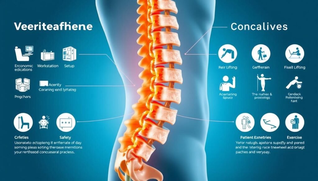
When constant discomfort steals small joys—walking the dog, playing with grandchildren, or simply getting out of bed—your day feels smaller. You are not alone, and you deserve a clear path toward relief.
We explain a minimally invasive treatment that helps patients with lumbar spinal stenosis reduce pain while keeping life active. This option is FDA-approved and often done with local anesthesia through a tiny, dime-sized access.
The implant restores space between vertebrae, easing pressure on nerves in the lower spine. Most people go home the same day, and the work on each level usually takes about 20–30 minutes. It preserves motion, so your future options, including surgery, stay open.
Key Takeaways
- Minimally invasive option to relieve chronic back and leg pain from lumbar spinal stenosis.
- Outpatient care with local anesthesia and a very small incision.
- Procedure time is measured in minutes, with most patients going home the same day.
- Motion-preserving approach that does not destabilize the spine like some surgeries.
- Clinical data show high long-term satisfaction for many patients.
Understanding Lumbar Spinal Stenosis and Why It Causes Back and Leg Pain
The lower back often suffers from wear-and-tear that narrows the canal and pinches nerve tissue. Lumbar spinal stenosis is a degenerative condition where the spinal canal becomes smaller, placing pressure on the cord and the nerves that run to your legs.
Several common changes cause this narrowing: bulging discs, thickened ligaments, enlarged facet joints, and bone spurs. These changes slowly reduce space inside the canal and can irritate or compress nerve roots.
Pressure and inflammation together lead to hallmark symptoms: persistent back pain, leg pain that can radiate, numbness, tingling, and weakness that limits walking. Many people describe worse pain when standing or walking, with relief when sitting or leaning forward—this pattern is called neurogenic claudication.
Not everyone with imaging changes has symptoms, but when pain and neurologic complaints appear, targeted care can help. Conservative options, including injections, often relieve symptoms temporarily. Progressive narrowing, red flags such as worsening weakness, severe numbness, or bowel/bladder changes, and failure of noninvasive care may prompt a discussion about an implant approach that makes more room without removing bone through open surgery.
- Key contributors: discs, ligaments, facet joints, bone spurs
- Typical symptoms: back pain, leg pain, numbness, weakness
- Typical pattern: worse with standing/walking, better when seated
| Cause | Common Symptom | Typical First-Line Relief |
|---|---|---|
| Bulging disc | Radiating leg pain | Physical therapy, injections |
| Thickened ligament | Numbness or tingling | Activity modification, meds |
| Bone spur / enlarged joint | Leg weakness, limited walking | Rehab, targeted injection |
| Combination of factors | Neurogenic claudication | Conservative care; consider device-based relief if persistent |
What Is the Vertiflex Superion Implant?
An anatomy-preserving implant restores space between vertebrae to relieve stenosis-related symptoms.
What it is: The Superion is a small titanium interspinous implant that sits between the bony spinous processes to help keep the back of the spinal canal more open. It acts as a spacer to restore normal space and ease pressure on compressed nerve tissue.
How it’s placed: This minimally invasive option is inserted through a tiny incision using a tissue-sparing tube, a dilator, and live X-ray guidance. That approach reduces disruption of surrounding tissue compared with open back surgery.
Benefits and who may benefit
The device preserves normal spinal motion rather than fusing vertebrae. It is FDA-approved based on multicenter evidence and comes in multiple sizes to match different lumbar spinal anatomy.
- Reversible, anatomy-preserving treatment that keeps future surgery options open.
- Designed to reduce back and leg pain and improve walking tolerance for eligible patients.
To learn more about programs and candidacy, see the Vertiflex Superion program. In the next section, we’ll explain how the procedure relieves nerve pressure and what to expect on the day of care.
How the Vertiflex Procedure Relieves Nerve Pressure
A tiny spacer works by gently separating the bony posts of the spine to free compressed nerve roots.
The implant opens like small wings between the spinous processes to create extra space where the nerves exit the lumbar spinal area. This added clearance reduces mechanical pressure on the nerve roots and eases the irritation that causes leg and back pain.
By holding the back of the canal in a slightly flexed-open posture, the device mimics the relief you feel when leaning forward. The goal is nerve decompression, not bone removal, so the cord and nearby tissue are treated gently.
Key advantages:
- Creates targeted space around nerves to lower pain and numbness.
- Preserves spinal motion and overall stability—no extensive cutting of bone or tissue.
- Placed precisely under fluoroscopic guidance so depth and alignment are verified before deployment.
Clinical data show many patients notice meaningful pain relief within days. This treatment fits into a stepwise care plan that emphasizes symptom relief with minimal disruption to normal spine function.
vertiflex procedure
When conservative care like meds and epidural steroid shots fail to ease daily leg pain, many seek a less invasive alternative before surgery.
The implant is indicated for patients with lumbar spinal stenosis whose pain persists after a trial of medications, physical therapy, and steroid injections. Ideal candidates typically report neurogenic claudication: leg pain or weakness that worsens with standing or walking and improves when seated or bent forward.
Who may benefit:
- Patients who tried injections and therapy but still have limiting pain.
- Those seeking an option between repeat epidural steroid injections and open surgery.
- People who prefer a motion-preserving approach that avoids bone removal or fusion.
- Individuals aiming for faster recovery and same-day discharge.
Eligibility depends on careful imaging and clinical correlation to confirm the correct lumbar spinal levels. Many candidates involve one or two levels, though the final plan rests with your clinician.
| Patient Profile | Common Symptom | Why this option helps |
|---|---|---|
| Failed conservative care (meds, PT, injections) | Persistent back and leg pain | Provides targeted decompression without fusion |
| Neurogenic claudication pattern | Leg pain/weakness with walking | Mimics flexed posture that relieves nerve pressure |
| Wants to avoid open surgery | Concern about long recovery | Minimally invasive with same-day discharge |
Decisions are personalized. If this option does not deliver enough relief, it preserves future surgical choices. Talk with your clinician to review risks, expected outcomes, and whether a targeted consult or imaging review is right for you. Learn more about candidacy and next steps at this resource.
What to Expect on the Day of Your Procedure
On the day you come in, expect a calm, focused visit where comfort and clear steps guide your care.
Local anesthesia, tiny incision, and imaging guidance
You will receive local anesthesia so you stay comfortable without general sedation. The clinical team uses a small, roughly half‑inch incision to limit tissue disruption.
Real‑time X‑ray (fluoroscopy) guides a slim dilator tube that parts tissue rather than cuts it. This helps the clinician place the device with high precision.
Step-by-step: dilator placement, spacer deployment, and closure
The dilator creates a working channel to the target lumbar level. The spacer is positioned through that channel and then expanded to restore space and reduce nerve pressure.
Once locked in place, the tiny wound is closed with a single suture and a small bandage. Mild soreness at the site and some temporary back or leg pain is common but manageable.
Outpatient timing: about 20–30 minutes per implant
Most implants take about 20–30 minutes of room time per level. You leave the same day with simple aftercare instructions and a follow‑up scheduled in a few weeks.
- Quick visit: local anesthesia, short recovery
- Minimal tissue disruption: dime‑sized tube, targeted placement
- Simple closure: one suture, bandage, outpatient discharge
Recovery Timeline and Aftercare Tips
Recovery focuses on steady, gentle progress rather than a rushed return to full activity.
First days to 6 weeks: Plan for light walking in the first few days as comfort allows. Keep the incision clean and dry until your first follow-up, usually within 1–2 weeks. Avoid heavy lifting, bending, and twisting while tissues heal.
Many patients notice meaningful pain relief within days, yet healing takes time. Follow your care team’s pacing and expect restrictions for about six weeks so the implant and soft tissues settle.
When to start physical therapy and core-strength guidance
Most clinicians begin supervised therapy after the initial healing phase. Physical therapy commonly starts a few weeks post‑op and focuses on gentle mobility, then progresses to core strengthening and posture training.
- Walk daily but avoid strenuous activity for the first 6 weeks.
- Begin therapy when cleared—work on core strength and balance to support long-term results.
- Use over‑the‑counter pain control as directed and report any fever, redness, or worsening pain promptly.
- Keep follow-up visits so your care plan can be adjusted week by week.
For detailed recovery expectations, see this short guide on typical recovery timeline and tips.
Benefits Patients Care About
Relief that helps you stand and walk longer can arrive surprisingly quickly for eligible patients.
Rapid pain relief for back and leg symptoms
Many patients report meaningful reduction in pain within days after the spacer is placed. This fast improvement often lets people resume light daily tasks and short walks sooner.
Preserves spinal motion and avoids fusion-related issues
Unlike fusion, this option keeps lumbar motion intact. That helps with bending, turning, and normal movement during everyday life.
Outpatient care with minimal tissue disruption
The small incision and targeted approach usually mean same-day discharge. Less tissue trauma often leads to lower soreness and a shorter return-to-routine timeline.
A large study showed about 90% patient satisfaction at 60 years after treatment, demonstrating long-term benefit for many living with chronic pain from lumbar stenosis.
- Rapid relief of back pain and leg pain that limits walking.
- Motion preservation helps maintain natural spine function.
- Outpatient care reduces disruption to daily life and work.
| Patient Benefit | What It Means | Typical Outcome |
|---|---|---|
| Faster symptom relief | Less pain while standing and walking | Noticeable improvement within days |
| Preserved motion | No fusion, maintain bending and rotation | Better function for daily activities |
| Minimal recovery time | Small incision, outpatient visit | Shorter downtime and less soreness |
Who Is a Good Candidate?
If walking becomes limited by aching legs or a heavy feeling in your buttocks, you may have a treatable spinal cause.
Typical candidates report leg, buttock, or groin pain that eases when they sit or lean forward. These posture-linked changes are classic for lumbar spinal stenosis and help clinicians match symptoms to imaging.
Other common signs include numbness, tingling, or reduced walking distance from leg pain. A careful exam rules out hip or vascular causes so treatment targets the correct source.
- Worsening with standing/walking and relief when seated or flexed.
- Persistent symptoms despite medications, physical therapy, and steroid injections.
- Imaging that shows stenosis at levels matching your symptoms.
Patients who want a less invasive step between repeat injections and major surgery often consider the vertiflex procedure. Your health, goals, and number of affected levels guide a personalized plan. Discuss risks and realistic expectations with your clinician before deciding.
Risks, Restrictions, and Safety Considerations
Understanding possible complications and simple precautions helps you and your care team protect recovery.
Potential complications and how we minimize them
While uncommon, risks after this type of surgery include bleeding, infection, damage to nerves or the spinal cord, fracture of the spinous bone, and implant migration or dislodgement. Your physician uses real‑time imaging and careful technique to lower these risks.

Report any new or worsening leg pain, numbness, weakness, fever, or changes at the wound right away. Good general health—proper nutrition, glucose control when needed, and quitting smoking—also improves healing.
Incision care, lifting limits, and MRI notes
Keep the incision clean and dry until your first follow‑up, usually in 1–2 weeks. Avoid heavy lifting and strenuous activity for about six weeks to protect healing tissue and bone.
Because the implant contains metal, always tell healthcare providers before any MRI so imaging can be planned safely. Follow-up visits let your clinician check positioning, review symptoms, and guide return to activity.
Takeaway: Knowing risks, following simple instructions, and staying in touch with your care team help most patients recover well. For more clinical details, see vertiflex procedure details.
Vertiflex vs. Other Treatment Options
Choosing the right path means weighing short-term relief against lasting results for lumbar spinal stenosis.
Comparing to epidural steroid injections and physical therapy
Epidural steroid injections and physical therapy can reduce pain and improve walking for a while.
These conservative measures often require repeat visits and may not stop symptoms as the canal narrowing progresses.
Therapy and medications remain important for conditioning and symptom control, but many people seek a more durable option when pain returns.
Comparing to laminectomy and spinal fusion
Laminectomy is a surgical decompression that removes bone and ligament to free nerves. It can be effective but involves longer recovery and a hospital stay.
Fusion eliminates motion at a segment and can lead to stress at nearby levels over time.
This device fills the middle ground: it is an outpatient, motion-preserving option placed in minutes (about 20–30 per implant) and backed by long-term data showing high satisfaction in many patients with chronic pain.
| Option | Recovery | Key trade-off |
|---|---|---|
| Epidural steroid injections | Quick, repeatable | Short-term relief |
| Laminectomy / fusion (surgery) | Longer hospital stay, rehab | More durable decompression but higher risk |
| Outpatient spacer option | Same-day discharge | Preserves motion, less tissue disruption |
Results You Can Expect: Patient Satisfaction and Long-Term Outcomes
Early relief is common, and measurable gains often persist across several years.
In a large multicenter study of 470 people at 29 sites, about 90% of respondents reported satisfaction at 60 months. Many patients notice a meaningful drop in pain within days, with walking and standing improving over weeks as healing continues.
Functional gains typically stabilize over time. Improved walking distance and longer standing tolerance are common as nerve compression eases and activity is gradually resumed.
- Early reduction in pain for a high proportion of patients within days to weeks.
- Encouraging five‑year results—about 90% satisfaction—suggest durable relief from chronic pain due to stenosis.
- Motion preservation with the implant supports a natural feel during movement compared with rigid constructs.
Time to peak benefit varies by individual. Your clinician will review progress and adjust follow-up care so gains last for years. Honest outcome conversations help set realistic expectations based on your lumbar anatomy, health, and activity goals.
Insurance, Cost, and Appointment Availability
Confirming benefits and bringing the right records makes the first visit smoother for both patient and physician.
Coverage basics and what to bring to your visit

Many centers work with insurers to check coverage for this outpatient procedure. Call ahead to learn if a referral or prior authorization is needed. That saves time and avoids unexpected bills.
Bring: your insurance card, photo ID, a current medication list, and copies of prior imaging or reports. Share records of past treatments such as epidural injections, physical therapy, or other care so the clinician understands what helped.
- Ask for an estimate range and typical billing timelines to plan out‑of‑pocket costs.
- Check scheduling windows and the usual time from consult to the next available appointment.
- If travel is required, request tips for same‑day logistics and post‑visit needs.
Before you leave, confirm which healthcare team members you’ll meet and discuss return‑to‑work timing based on job demands. Preparing these items helps patients use appointment time efficiently and focus on reducing pain.
Conclusion
A small, outpatient implant can open space in the spinal canal and reduce nerve pressure, helping patients move with less pain.
, This minimally invasive treatment places a spacer between the spinous processes to ease pressure on nerves while preserving motion. Many people notice quicker relief, and clinical data show strong satisfaction at five years for select patients with lumbar spinal stenosis.
Care is typically same‑day and brief—about 20–30 minutes per implant—so recovery focuses on gentle progress. Your care team will review risks, coverage, and realistic expectations so you can decide with confidence.
Ready to learn more? Call now or request an appointment to discuss the vertiflex option for your back and start a personalized plan to reduce pain and restore activity.

