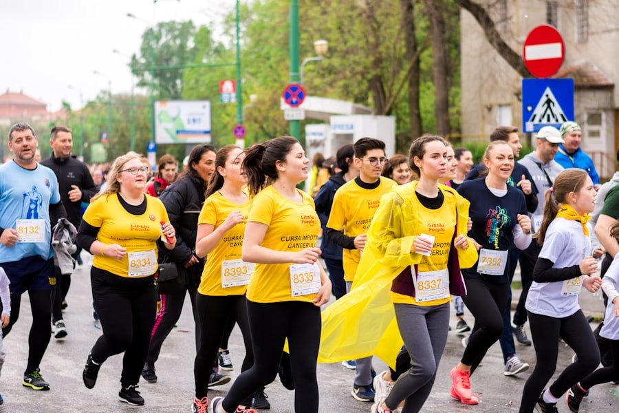
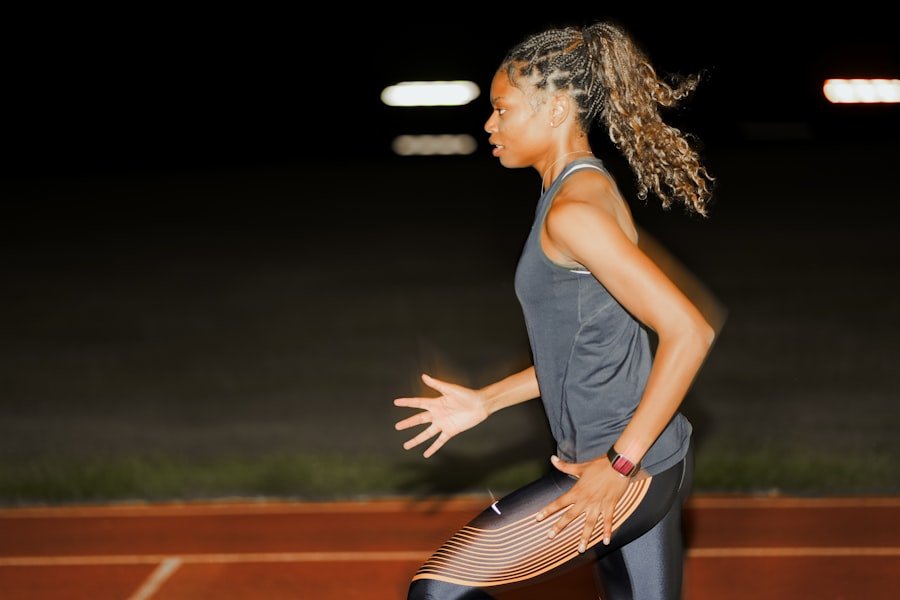
Iliotibial Band Syndrome (ITBS) is a common overuse injury that primarily affects runners, cyclists, and individuals engaged in repetitive knee-bending activities. This condition arises when the iliotibial band, a thick band of connective tissue that runs along the outside of the thigh from the hip to the shin, becomes tight or inflamed. The syndrome is characterized by pain on the outer side of the knee, which can significantly hinder physical activity and diminish quality of life.
Understanding ITBS is crucial for athletes and active individuals, as it can lead to prolonged discomfort and may require extensive rehabilitation if not addressed promptly. The prevalence of ITBS has garnered attention in sports medicine, particularly due to its impact on performance and training regimens. Athletes often push their limits, leading to biomechanical imbalances that can exacerbate this condition.
As such, recognizing the signs and symptoms early on is essential for effective management. This article delves into the anatomy of the iliotibial band, explores the causes and symptoms of ITBS, discusses diagnostic methods, and outlines treatment options, prevention strategies, and rehabilitation exercises.
Key Takeaways
- I. Introduction to Iliotibial Band Syndrome
- Iliotibial Band Syndrome is a common overuse injury that causes pain on the outer side of the knee, often experienced by runners and cyclists.
- II. Anatomy of the Iliotibial Band
- The iliotibial band is a thick band of fascia that runs along the outside of the thigh, connecting the hip and the knee.
- III. Causes of Iliotibial Band Syndrome
- Overuse, muscle imbalances, improper training techniques, and biomechanical issues can contribute to the development of Iliotibial Band Syndrome.
- IV. Symptoms of Iliotibial Band Syndrome
- Symptoms include pain on the outside of the knee, swelling, and a clicking or popping sensation during movement.
- V. Diagnosis of Iliotibial Band Syndrome
- Diagnosis is typically based on a physical examination, medical history, and imaging tests such as MRI or ultrasound.
- VI. Treatment Options for Iliotibial Band Syndrome
- Treatment may include rest, ice, physical therapy, and in severe cases, surgery to release the tight band of tissue.
- VII. Prevention of Iliotibial Band Syndrome
- Prevention strategies include proper warm-up, stretching, gradual increase in training intensity, and addressing any biomechanical issues.
- VIII. Exercises for Iliotibial Band Syndrome
- Strengthening and stretching exercises for the hip and thigh muscles can help prevent and rehabilitate Iliotibial Band Syndrome.
- IX. Recovery and Rehabilitation for Iliotibial Band Syndrome
- Recovery may involve a gradual return to activity, continued physical therapy, and addressing any underlying causes of the syndrome.
- X. Complications of Iliotibial Band Syndrome
- If left untreated, Iliotibial Band Syndrome can lead to chronic pain and potentially affect an individual’s ability to participate in physical activities.
- XI. Conclusion and Future Outlook for Iliotibial Band Syndrome
- With proper diagnosis, treatment, and prevention strategies, individuals can effectively manage Iliotibial Band Syndrome and continue to engage in their desired physical activities.
Anatomy of the Iliotibial Band
The iliotibial band is a fibrous structure that plays a vital role in stabilizing the knee during movement. It originates from the tensor fasciae latae muscle and the gluteus maximus muscle, extending down the lateral aspect of the thigh to insert at the lateral condyle of the tibia.
This band serves as an important stabilizer for the knee joint, particularly during activities that involve running or cycling.
Its primary function is to assist in maintaining proper alignment of the knee while allowing for flexion and extension. The iliotibial band is not merely a passive structure; it interacts dynamically with surrounding muscles and tendons. The gluteus maximus and tensor fasciae latae muscles work in concert with the iliotibial band to control hip movement and stabilize the pelvis during locomotion.
When these muscles are weak or imbalanced, it can lead to increased tension on the iliotibial band, contributing to inflammation and pain. Understanding this anatomy is crucial for identifying risk factors associated with ITBS and developing effective treatment strategies.
Causes of Iliotibial Band Syndrome
Several factors contribute to the development of Iliotibial Band Syndrome, with overuse being the most significant. Activities that involve repetitive knee flexion and extension, such as running or cycling, can lead to microtrauma in the iliotibial band. This repetitive strain can cause irritation at the point where the band crosses over the lateral femoral epicondyle, resulting in inflammation and pain.
Additionally, training errors such as sudden increases in mileage or intensity can exacerbate these issues. Biomechanical factors also play a critical role in the onset of ITBS. Individuals with poor lower limb alignment, such as excessive pronation or supination of the foot, may place additional stress on the iliotibial band during movement.
Furthermore, muscle imbalances—particularly weakness in the hip abductors or external rotators—can lead to altered gait mechanics that increase tension on the band. Other contributing factors include inadequate warm-up routines, improper footwear, and uneven surfaces that can further strain this already vulnerable structure.
Symptoms of Iliotibial Band Syndrome
The hallmark symptom of Iliotibial Band Syndrome is lateral knee pain, which typically manifests during physical activity. Athletes may initially experience discomfort that subsides with rest but can progress to persistent pain that interferes with daily activities. The pain often intensifies during activities such as running downhill or cycling, where increased flexion of the knee places additional strain on the iliotibial band.
In some cases, individuals may also report a clicking or snapping sensation as the band moves over bony structures. In addition to pain, swelling may occur around the lateral aspect of the knee due to inflammation. This swelling can lead to stiffness in the joint, making it difficult for individuals to fully extend or flex their knee without discomfort.
As symptoms worsen, individuals may find themselves limping or altering their gait to avoid pain, which can further exacerbate underlying biomechanical issues and lead to additional injuries.
Diagnosis of Iliotibial Band Syndrome
Diagnosing Iliotibial Band Syndrome typically involves a thorough clinical evaluation by a healthcare professional. The process begins with a detailed medical history that includes information about symptoms, activity levels, and any previous injuries. A physical examination follows, during which a clinician assesses range of motion, strength, and any areas of tenderness along the iliotibial band and lateral knee.
One common diagnostic test is the Ober’s test, which evaluates tightness in the iliotibial band by assessing hip adduction range of motion. If the leg does not drop below horizontal when released from a flexed position, it may indicate tightness in the band. Imaging studies such as MRI or ultrasound are not typically required for diagnosis but may be utilized in complex cases to rule out other conditions such as meniscal tears or bursitis.
Treatment Options for Iliotibial Band Syndrome
Treatment for Iliotibial Band Syndrome often begins with conservative measures aimed at reducing inflammation and alleviating pain. Rest is crucial; individuals are advised to temporarily cease activities that exacerbate symptoms while allowing time for healing. Ice therapy can be beneficial in managing inflammation and should be applied to the affected area for 15-20 minutes several times a day.
Physical therapy plays a pivotal role in recovery from ITBS. A physical therapist will typically design a personalized rehabilitation program that includes stretching exercises to improve flexibility in the iliotibial band and surrounding muscles. Strengthening exercises targeting hip abductors and external rotators are also essential for restoring balance and stability to the lower extremities.
In some cases, corticosteroid injections may be considered for individuals who do not respond adequately to conservative treatments.
Prevention of Iliotibial Band Syndrome
Preventing Iliotibial Band Syndrome involves addressing risk factors before they lead to injury. One effective strategy is to incorporate a well-rounded training program that includes strength training, flexibility exercises, and proper warm-up routines before engaging in physical activity. Strengthening exercises for hip muscles can help maintain proper alignment during movement and reduce strain on the iliotibial band.
Footwear also plays a significant role in prevention; individuals should select shoes that provide adequate support and cushioning based on their foot type and gait mechanics. Additionally, gradually increasing training intensity and mileage can help prevent overuse injuries by allowing time for adaptation. Cross-training with low-impact activities such as swimming or cycling can also reduce repetitive stress on the iliotibial band while maintaining cardiovascular fitness.
Exercises for Iliotibial Band Syndrome
A targeted exercise regimen is essential for both rehabilitation and prevention of Iliotibial Band Syndrome.
Stretching exercises focusing on the iliotibial band itself as well as surrounding muscle groups are crucial for improving flexibility.
One effective stretch involves standing with one leg crossed behind the other while leaning towards the side of the back leg; this helps elongate the iliotibial band.
Strengthening exercises should focus on hip abductors and external rotators to enhance stability during movement. Clamshells—performed by lying on one side with knees bent and lifting the top knee while keeping feet together—are particularly effective for targeting these muscle groups. Additionally, single-leg squats can help improve overall lower limb strength while promoting proper alignment.
Recovery and Rehabilitation for Iliotibial Band Syndrome
Recovery from Iliotibial Band Syndrome varies depending on individual circumstances but generally involves a gradual return to activity following a structured rehabilitation program. Initially, individuals should focus on reducing pain and inflammation through rest and ice therapy before progressing to stretching and strengthening exercises under professional guidance. As symptoms improve, individuals can begin reintroducing low-impact activities such as swimming or cycling before transitioning back to running or other high-impact sports.
Monitoring symptoms closely during this phase is essential; if pain recurs, it may indicate that further modifications are needed in training intensity or technique. Regular follow-ups with healthcare professionals can help ensure that recovery remains on track.
Complications of Iliotibial Band Syndrome
While many individuals recover fully from Iliotibial Band Syndrome with appropriate treatment, complications can arise if left untreated or improperly managed. Chronic pain may develop if inflammation persists over time, leading to compensatory movement patterns that increase the risk of additional injuries elsewhere in the body—such as patellar tendinitis or hip bursitis. In some cases, persistent ITBS may require more invasive interventions such as surgical release of the iliotibial band if conservative measures fail after an extended period.
This option is typically reserved for severe cases where quality of life is significantly impacted by ongoing pain and dysfunction.
Conclusion and Future Outlook for Iliotibial Band Syndrome
As awareness of Iliotibial Band Syndrome continues to grow within both athletic communities and healthcare settings, ongoing research into its causes, treatment options, and prevention strategies remains vital. Advances in biomechanics and sports medicine are likely to yield new insights into effective management techniques that can enhance recovery outcomes for affected individuals. Future studies may focus on identifying specific risk factors associated with ITBS across different populations or exploring innovative rehabilitation methods that incorporate technology such as wearable devices for real-time feedback on movement patterns.
By fostering a deeper understanding of this condition, healthcare professionals can better equip athletes and active individuals with tools necessary for preventing injury while optimizing performance.
If you are interested in learning more about the physiology and psychology of pain, I recommend checking out the article Why Do We Feel Pain? Physiology and Psychology of Pain. Understanding the mechanisms behind pain can provide valuable insights into conditions like Iliotibial Band Syndrome and how to effectively manage them.
FAQs
What is Iliotibial Band Syndrome?
Iliotibial Band Syndrome (ITBS) is a common overuse injury that affects the iliotibial band, a thick band of fibrous tissue that runs along the outside of the thigh, from the hip to the knee.
What are the symptoms of Iliotibial Band Syndrome?
Symptoms of ITBS may include pain on the outside of the knee, swelling, aching or burning sensations, and a clicking or popping sensation when bending the knee.
What causes Iliotibial Band Syndrome?
ITBS is often caused by repetitive friction of the iliotibial band against the outside of the knee during activities such as running, cycling, or hiking. Other contributing factors may include muscle imbalances, overtraining, or improper footwear.
How is Iliotibial Band Syndrome diagnosed?
ITBS is typically diagnosed based on a physical examination and a review of the individual’s medical history. Imaging tests such as MRI or ultrasound may be used to rule out other potential causes of the symptoms.
What are the treatment options for Iliotibial Band Syndrome?
Treatment for ITBS may include rest, ice, compression, and elevation (RICE), as well as stretching and strengthening exercises, physical therapy, and the use of orthotics or supportive footwear. In some cases, corticosteroid injections or surgery may be necessary.